BioREE™ product series is designed to expand the possibilities of scanning electron microscopy (SEM) in analyzing various biological samples as well as to ensure prolonged preservation of the latter for subsequent molecular analysis.
Report on BioREE™ at the XX International Scientific Conference “Stochikovsky Readings”

Delicate preparation
for visualization

Enhanced contrast
and fixation

Preservation for
molecular analysis
We are offering you a completely new method of sample preparation that has the following benefits:
- Less time-consuming sample preparation for SEM
- No necessity for hard sample fixation, possibility of working with delicate objects
- Non-toxic and easy-to-use reagents
- More informative results, with no analogues in the application area of SEM
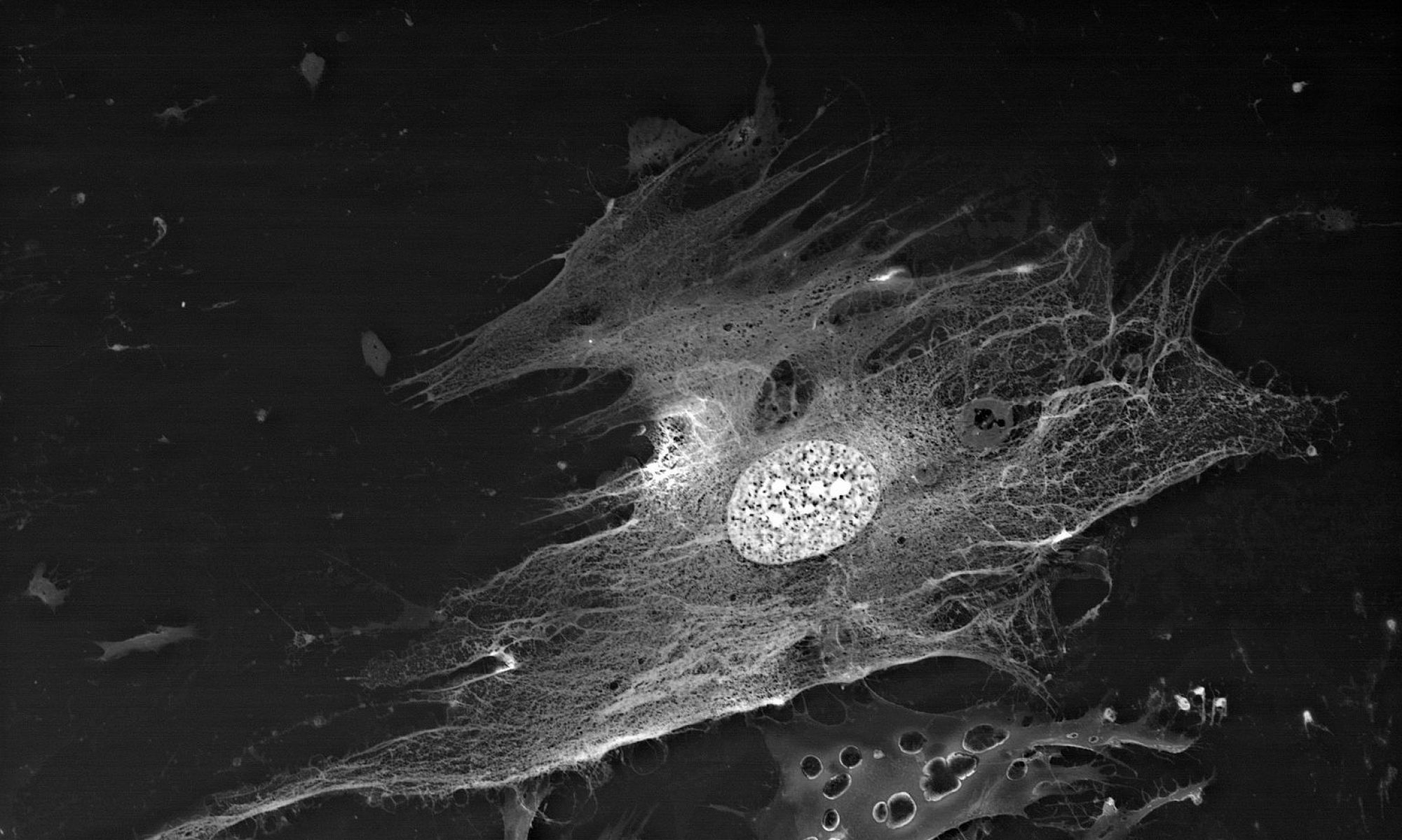
The BioREE technology is based on supravital staining with lanthanoid metal ions (La3+, Nd3+, Sm3+, etc.).By invading the cell metabolism and inhibiting its metabolic pathways, heavy lanthanoid cations mark the internal structure of the cell making it visible for SEM.
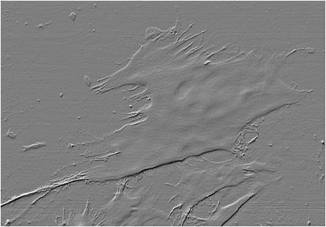
Classic SEM
(high vacuum, Pd/Au coating, SE mode)
Lanthanoid staining
(low vacuum, pretreatment with the «BioREE» set, BSE mode)
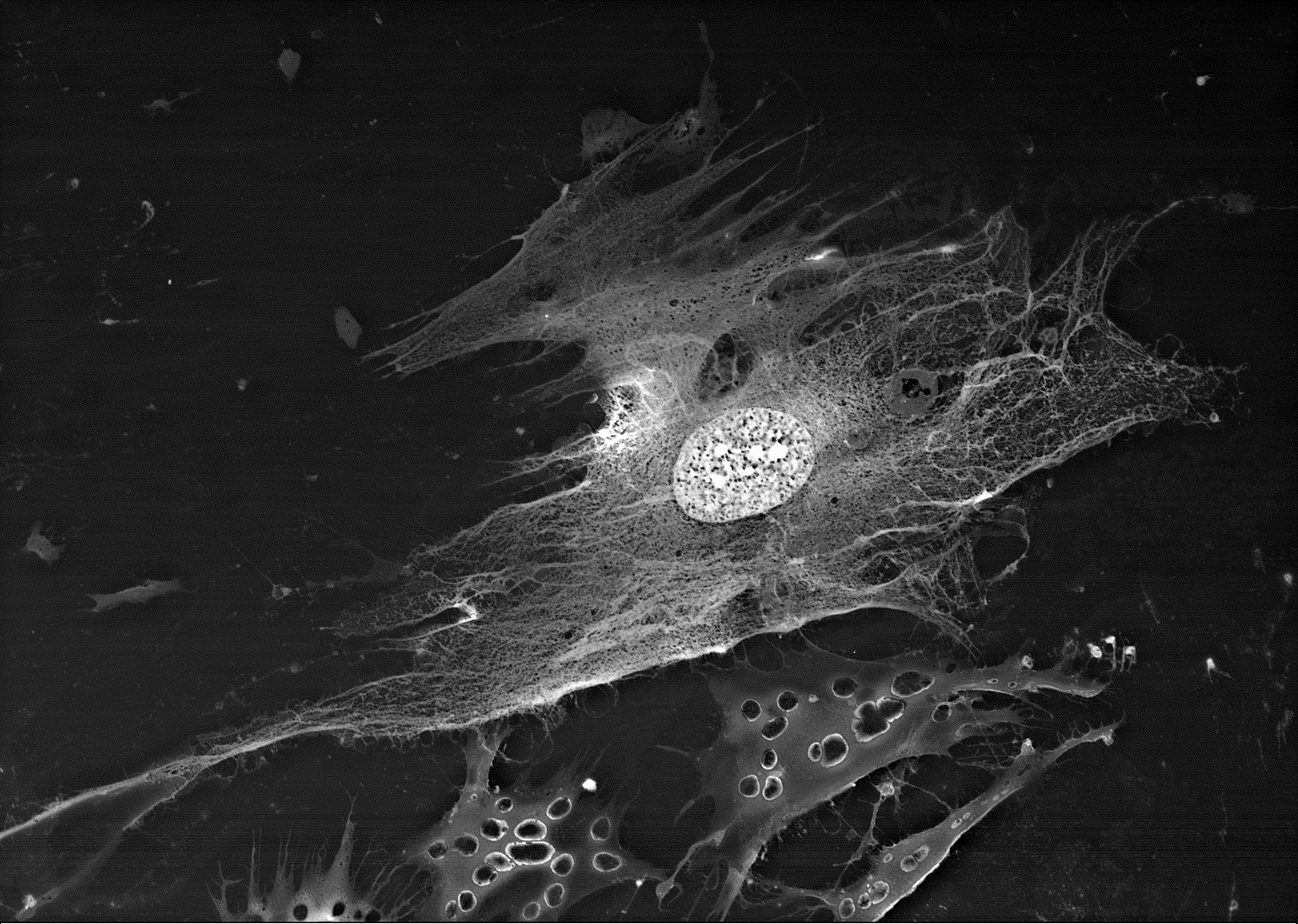
MMSC (mesenchymal stem cells) on cell culture plastic
Unlike the classical sample preparation for SEM, our methodology doesn’t use: osmium tetroxide, osmium thiocarbohydrazide, uranyl acetate, glutaraldehyde, paraformaldehyde
Lead-salt supplemented lanthanoid staining
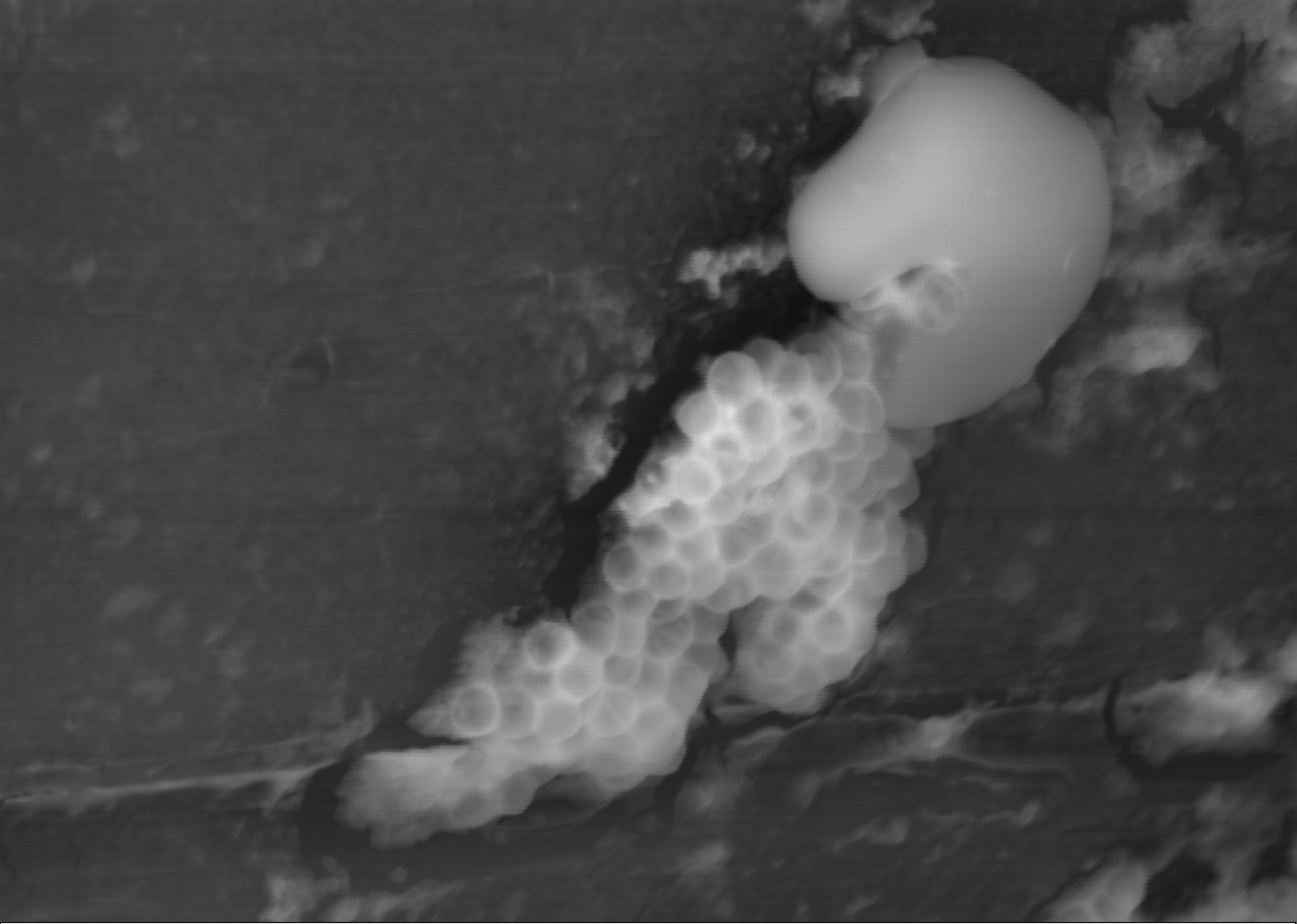
(low vacuum, pretreatment with the «BioREE—B» set, BSE mode)
Staining process
BioREE™ dramatically expands SEM possibilities of studying various biological objects as compared to common methods of sample preparation:
- High-resolution visualization of subsurface ultrastructure
- Easier cell identification in complex objects and on different substrates due to higher contrast of the sample
- Comparative analysis of functional condition of sample cells
- A more precise chemical microanalysis of the sample due to the absence of electroconductive coating
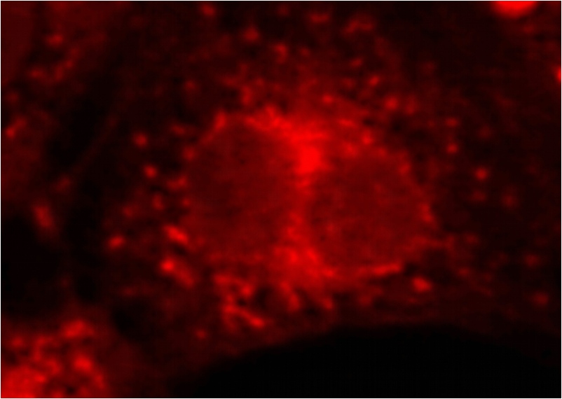
Ultrahigh-resolution fluorescent microscopy systems can be called the closest analogues of the BioREE-facilitated SEM in terms of the level of visualization. However, our method is:
- -more versatile and does not require the use of multiple specific dyes for visualization of different cell structures;
- -provides a significantly greater depth of field of the resulting images without compromising their resolution.
Because of the increasing popularity of correlative microscopy, the method of lanthanoid staining gains additional advantages, such as a higher survival of fluorescent marks and minimal deformation of the sample during its pretreatment, which allows for a more precise matching of images obtained by different methods.
Fluorescent staining
Lanthanoid staining
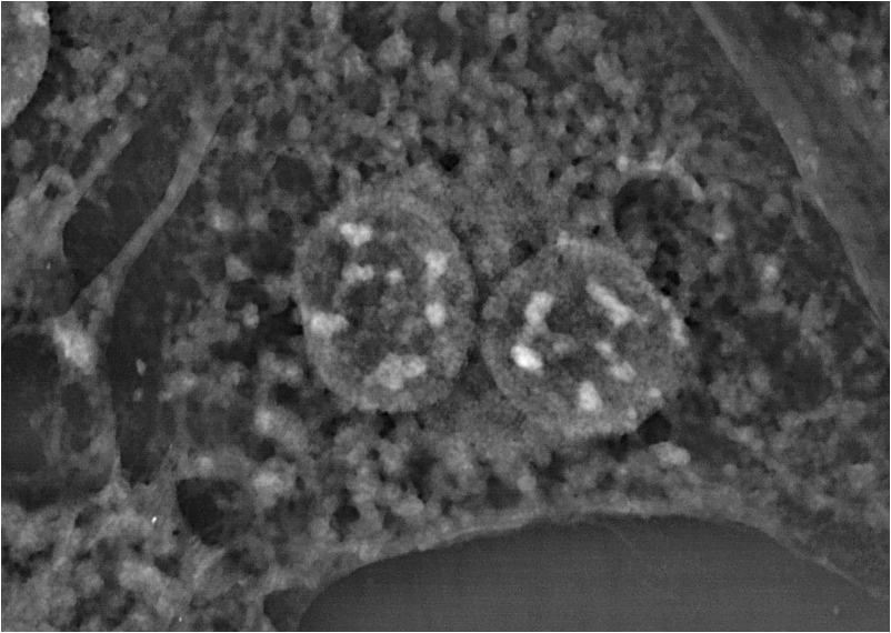
Mitochondria in human astrocytes